mri b value
To evaluate the performance of computed high b value diffusion-weighted images DWI in prostate cancer detection. The degree of diffusion weighting correlates with the strength of the diffusion gradients characterized by the b-value which is a function of the gradient related parameters.

Diffusion Weighted Imaging In Acute Ischemic Stroke Radiology Reference Article Radiopaedia Org
97 consecutive patients who had undergone multiparametric MRI of the prostate.
. For example consider series 16 from this archive. Our purpose was to evaluate the appearance of the normal brain on DW MR images as the diffusion gradient strength b value is increased from 1000 to 3000 smm2. A b value of 8001000 smm 2 would provide an excellent spatial resolution and an adequate signalnoise ratio for lesion evaluation.
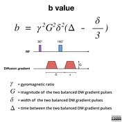
Mean ADC value is 13 higher in total by additional use of b 0 and b 50 smm 2 in multiple b -value combinations. B γ² G² δ² Δδ3 Therefore a larger b value is achieved by increasing the gradient amplitude and duration and by widening the interval between paired. As MR scanner hardware has improved allowing for increased gradient strengths we are able to generate higher b values for diffusion-weighted DW imaging.
8 reported b 3200 smm 2 was optimal among b values of 20004500 smm 2. Since 2017 7 T clinical scanners have been available see ultrahigh field MRI. The B 0 in MRI refers to the main static magnetic field and is measured in teslas TThe majority of MRI systems in clinical use are 15 T with increasing numbers of 3 T systems being installed.
With b 0 bright signals are noted in multiple veins due to the high T2 of blood coupled with sluggish flow. Certain illnesses show restrictions of diffusion for example demyelinization and cytotoxic edema. The lesionnormal parenchymal ADC ratio for b 600 b 1000 and multiple b 2 better distinguished between benign and malignant lesions.
Higher b values typically range between 500 and 1000 smm 2. Mean ADC values were significantly different between malignant and benign lesions for all b-value combinations P0000. In contrast this molecular motion may be obstructed in certain pathological conditions - such as in the.
This MR technique detects diffusion abnormalities which can be quantified by computing apparent diffusion coefficient ADC maps. B value measures the degree of diffusion weighting applied thereby indicating the amplitude G time of applied gradients δ and duration between the paired gradients Δ and is calculated as. Two recent studies explored DWI at an ultra high b-value of 2000smm 2.
Thirty-five PCa patients who were to be treated with radical prostatectomy underwent 3T DWI. 2015 designed a study to correlate the accuracy of 3T MRI in which DWI occurred with a b-value of 2000smm 2. Altering the field strength will affect the Larmor frequency at which the protons precess.
An in vivo liver mri results in the normal controls revealed that the measured pseudodiffusion value was lower when calculated with less low b values no b values between 0 and 50 smm 2 compared to when calculated with more low b value extra b 10 and 25 smm 2 were included in the low b-value distribution 41 which were consistent with. However there is no agreement concerning the number of images needed for ADC calculation. Guidance for Adjusting MRI Scan Sequence SAR and B1rms Values 2 MRI scanner operator andor radiologists are responsible for verifying compliance with all scan conditions identified in the product labeling.
Depending on the organ being imaged b-values typically range from 50-1000smm 2. Using lower b-values such as 0 and 50 together with higher b-values 600 smm2 was beneficial Az0928 and 0927. 2015 designed a study to correlate the accuracy of 3T MRI in which DWI occurred with a b-value of 2000smm 2.
This array of multiple white dots on the b0 image makes distinguishing cysts masses and vessels difficult. This series has images with b_values of both 750 and 1500 but the DICOM tag stores b_values of 1000001500 and 1000000750 as shown in the DICOM dump from image 24 below. The reason is readily apparent from the images below.
051-067 and a pooled specificity of 050 95 CI. The Specific Absorption Rate SAR is a measurement of RF energy deposition in the body expressed in watts per kilogram Wkg. The term b -value characterizes the diffusion gradient pulses amplitude shape and timing and expresses the amount of diffusion weighting 1.
ADC values are typically calculated from a set of MR images obtained with varying degrees of diffusion weighting b-values using nonlinear regression. Be aware that the b_value stored in tag 00431039 may be masked. DWI is done to determine the rate of molecular diffusion in different areas of the body.
Multiple b values of 600 smm 2 and higher are recommended to differentiate between benign and malignant abdominal lesions. From fast-moving to restricted hindered and slow-moving water molecules. The b value is used in MRI in the context of Diffusion Weighted Imaging DWI.
A baseline b-value of 50 smm² is often used in liver diffusion-weighted imaging instead of b 0. In DWI we recommend the use of b -values of 0 and 800 smm 2 as two b -values or b 0 50 600 800 and 1000 smm 2 as multiple b -values for distinguishing between benign and malignant liver lesions. The best b-value combination was 0 and 800 Az0935.
In biological tissues different regimes of water diffusion are encountered. Deliveries in some areas may be delayed due to current weather conditions nationwide. Therefore if you use 00431039 to determine b_value you.
The use of b values more than 1000 smm 2 would offer better contrast but was more liable to suffer susceptibility artifact. Conclusion In DWI the optimal b value is 600 smm 2. Strength duration and the period between diffusion gradients.
In general in healthy tissue molecules of water and other chemicals are not stationary but moving about. Mean ADC values were approximately 13 1-15 higher in.

Diffusion Tensor Imaging Dti Fiber Tracking Imagilys
2

Apparent Diffusion Coefficient Radiology Reference Article Radiopaedia Org

Principles Of Diffusion Tensor Imaging And Its Applications To Basic Neuroscience Research Neuron

Pulse Sequences For Diffusion Weighted Mri Sciencedirect
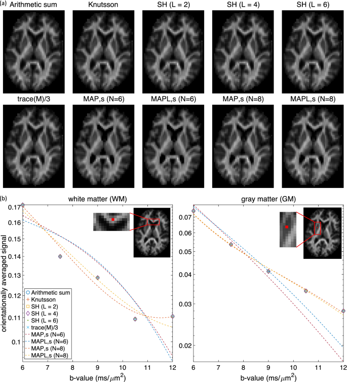
Computing The Orientational Average Of Diffusion Weighted Mri Signals A Comparison Of Different Techniques Scientific Reports
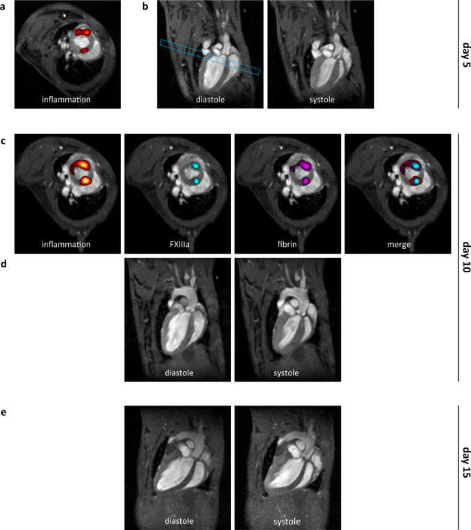
Multi Targeted 1h 19f Mri Unmasks Specific Danger Patterns For Emerging Cardiovascular Disorders Nature Communications
Diffusion Weighted Imaging Radiology Reference Article Radiopaedia Org
2

Hyperpolarised 13c Mri Identifies The Emergence Of A Glycolytic Cell Population Within Intermediate Risk Human Prostate Cancer Nature Communications
Tensor Valued Diffusion Encoding For Diffusional Variance Decomposition Divide Technical Feasibility In Clinical Mri Systems Plos One
Comparison Of 3d Printed Prostate Models With Standard Radiological Information To Aid Understanding Of The Precise Location Of Prostate Cancer A Construct Validation Study Plos One

Pulse Sequences For Diffusion Weighted Mri Sciencedirect

Diffusion Tensor Imaging Dti Fiber Tracking Imagilys
Diffusion Weighted Imaging Radiology Reference Article Radiopaedia Org

Apparent Diffusion Coefficient Radiology Reference Article Radiopaedia Org
Tensor Valued Diffusion Encoding For Diffusional Variance Decomposition Divide Technical Feasibility In Clinical Mri Systems Plos One
2

Diffusion Weighted Imaging Radiology Reference Article Radiopaedia Org
0 Response to "mri b value"
Post a Comment